Airway Simulator
4D printing | Medical design
Simulating patient pathology in an interactive trachea model for airway management training
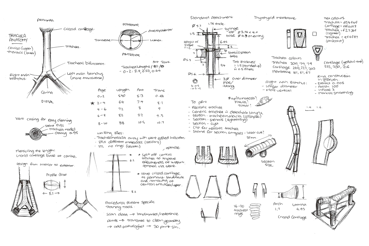
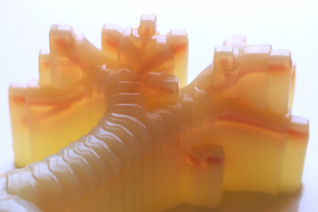
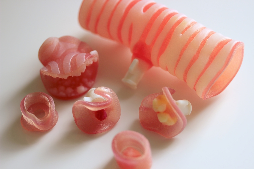
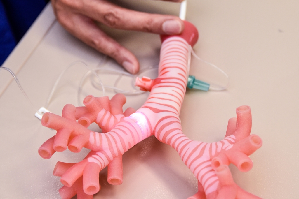
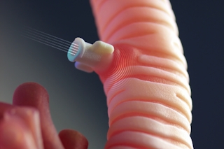
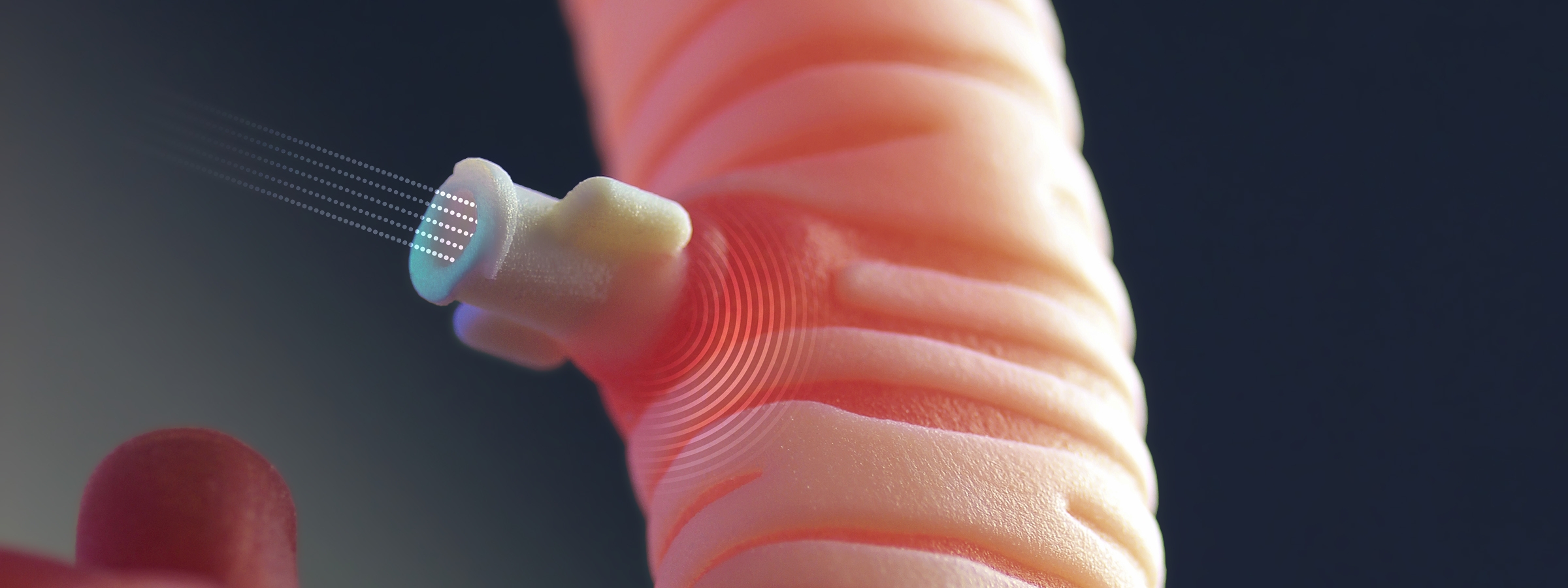
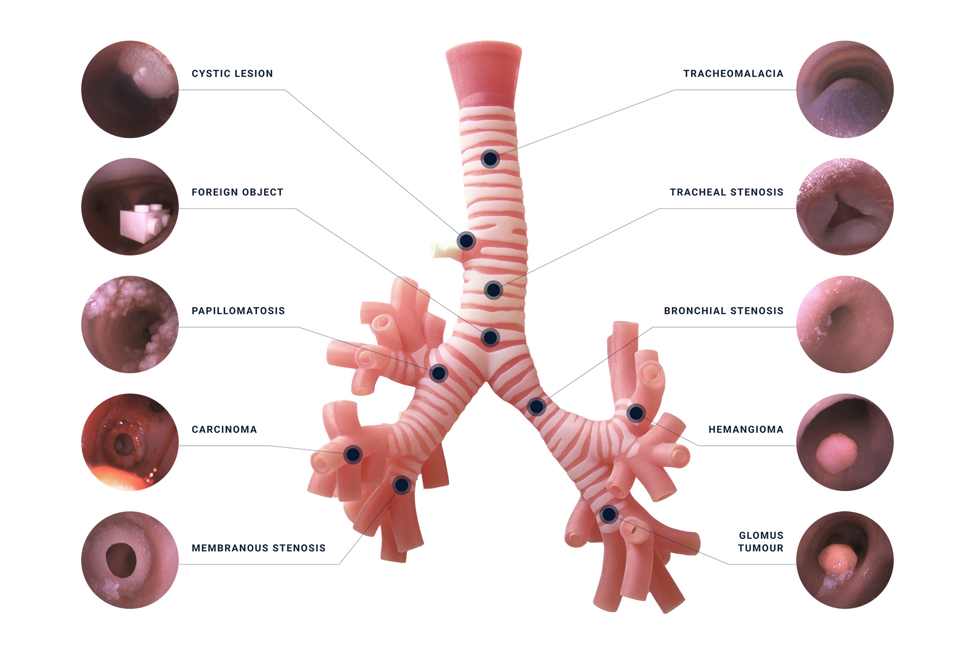
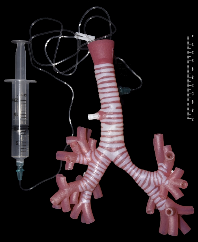
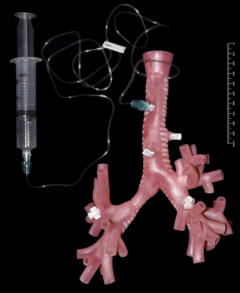
This 3D printed model of an adult trachea mimics the look, feel and function of complex human anatomy. It is designed for simulation purposes to give medical practitioners more realistic tactile and visual feedback. Novel dynamic parts with pneumatic and fluid mechanics feature throughout the trachea. Movements like fluid secretion, bleeding or constriction can be reproduced by attaching syringes. This interactive trachea allows abnormalities to be identified and procedures to be trained with a high degree of realism. There is also the opportunity to customise the model to patient-specific data.
Design Tools
3D Slicer, ZBrush, Rhino + Grasshopper, Houdini, Netfabb, GrabCAD, Stratasys J750 3D Printer, InDesign
My Role
Conceptualisation, 3D modelling, manufacture
Attribution
Created by the team at Simpath: Bernard Guy, James Broadbent, Jeremy Young, Nicole Hone
Medical evaluation
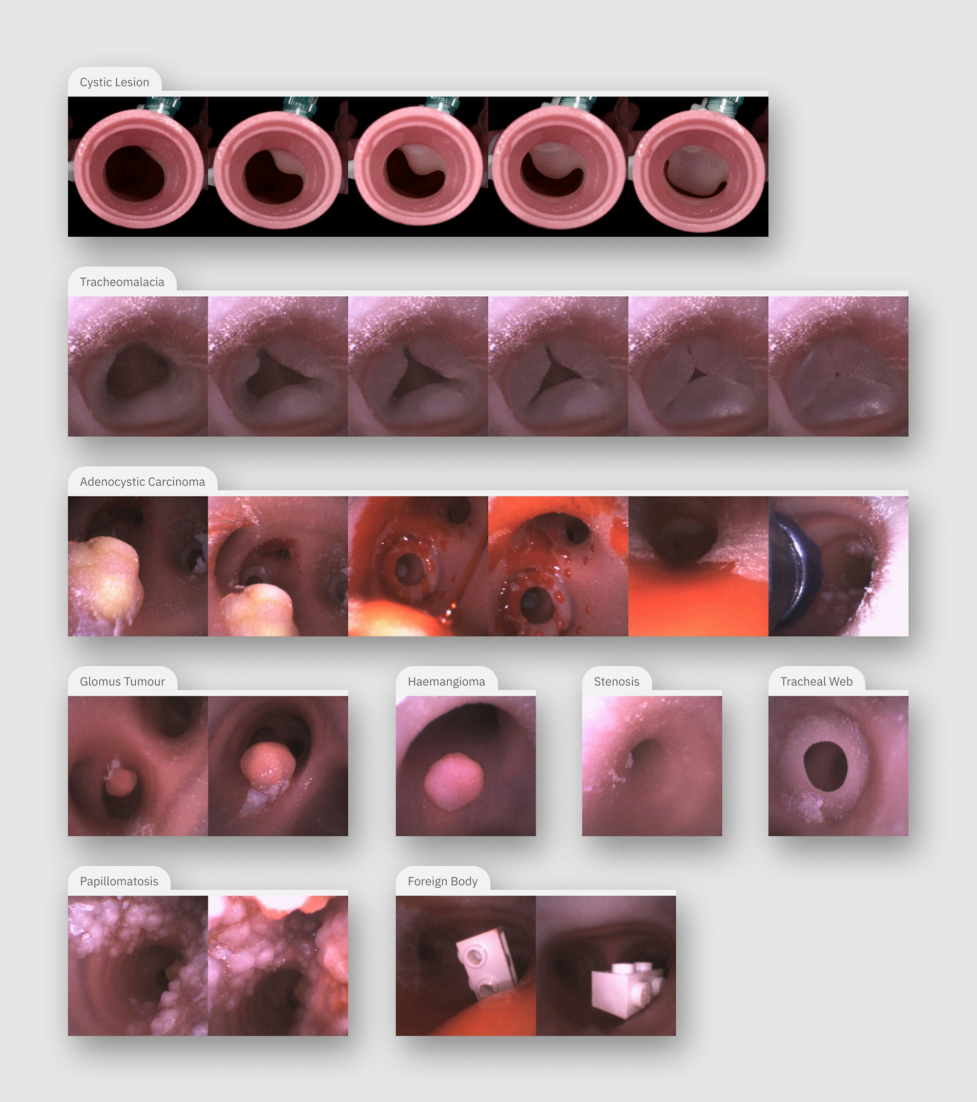
A team of anaesthetists tested the model in a simulated bronchoscopy. The sequences of images below capture the journey of the scope travelling through the model. The feedback loop between clinician and designer is vital to developing greater anatomical realism while ensuring the model can tolerate medical procedures.
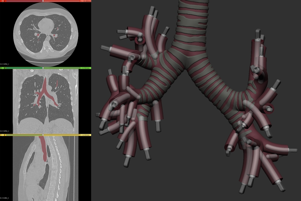
From scan to physical simulation
The trachea 3D model is an amalgamation of human scan data and custom-designed pathology. Firstly, the trachea is segmented from a scan of the thoracic region. The mesh is cleaned and anatomical details are restored. Careful research into medical reports, videos and images of airway pathology guide the design of abnormal features. Variable colour and material qualities are seen throughout the model thanks to digitally mixed PolyJet materials. This allows a smooth integration of firm cartilage rings through to soft bronchial endings.